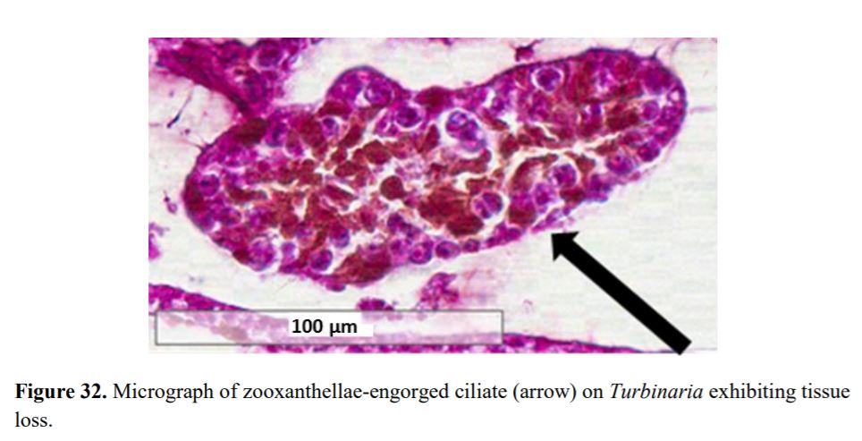
Protozoan is an informal term for single-celled eukaryotes, either free-living or parasitic, which feed on organic matter such as other microorganisms or organic tissues and debris. In this case, these protozoans feed on the zooxanthellae found in coral tissue causing the coral to tissue necrosis. They also feed on other protozoans cannibalizing each other.
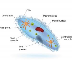
Protozoan is a single cell organism whose cell has a nucleus enclosed within a membrane. They possess animal-like behavior such as motility and predation. Protozoan range from 50 to 150 μm (micrometers) in with and 50 to 500 μm in length. Protozoa can reproduce sexually or asexually. Asexual reproduction is the process in which an organism produces offspring by itself, without the participation of another organism of its species. It does this by budding, a process where a new organism develops from an outgrowth or by cell division.
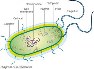
Bacteria is a single cell organism that has a cell wall but lack organelles and an organized nucleus. Most bacteria range from 0.2–2.0 µm (micrometers) in diameter. Less than 1 micron (0.001 mm/0.00004 inch) in length. Bacteria reproduces by binary fission, a process where a single cell divides into two identical daughter cells.